


The Tissue: Issue #2
Sharing the world’s first kidney transplant using a genetically-edited pig organ and commentary on the future of organ transplantation

12-minute read
Hello enthusiasts for living materials of all sorts!
A lot has happened in the field of tissue engineering over the last month, so I’m not going to waste any time on additional pleasantries, although I hope April treated you kindly. On March 26th, ARPA-H announced $190 million in funding for the development of transformative osteoarthritis therapies in what may be the single biggest public investment for the field. We’ll be talking in more detail about that announcement next month, but I couldn’t keep it from you until then!
On a more administrative note: I want to make The Tissue a space where we can feature Q&As, similar to the ones I’ve done with Dr. James Henstock and Corinne Okada Takara (both of which I recommend you read!), but in a more condensed format. Let me know in the comments if there’s anyone you’re interested in hearing from or any questions you’re interested in finding answers to!
New and Interesting Publications
How to commercialize regenerative medicine therapies: successful case studies
Published less than a year ago in September, 2023, this paper is a must read for any researchers who eventually want to see their research on regenerative therapies reach patients! In the paper, the authors outline the path to FDA-approval, providing case studies of products which successfully overcame barriers to translation, including Staratech’s StrataGraft, AveXis’s Zolgensma, and Spark Therapeutic’s Luxturna. They lay out many of the challenges and decisions researchers must face and make when translating scientific findings from the lab to the market, including how and when to apply for patents to establish intellectual property rights, what funding mechanisms to pursue to most effectively develop the required business and infrastructure, and how to navigate the regulatory pathway to successfully receive approval. There is some tension here, as noted by the authors. While commercialization requires “careful consideration of data ownership” and disclosure, the NIH as well as The White House are increasingly requiring the results of taxpayer-supported research to be publicly disclosed.
Periosteum-mimicking biomaterial successfully heals large bone defects by modulating bone, vascular, and immune cell survival and differentiation
In our never-ending quest as a field to more effectively regenerate bone, we often stumble down complicated rabbit holes. As many of you know, bone is a tissue that continues to remodel throughout our lives, subject to the whims of osteoclasts and osteoblasts absorbing and depositing new mineral in a never-ending cycle. Much of bone’s ability to heal comes from the periosteum, the sheath of blood vessels, nerves, and immune cells that enshroud it and activate when injured. In this study, researchers developed a “hamburger-like nutrient periosteum structure (HNS)” to mimic the role of periosteum in bone healing. Their scaffold—a central cell sheet composed of bone-marrow-derived mesenchymal stromal cells and M2 macrophages sandwiched between a silk fibroin hydrogel containing PLLA microspheres and CaO2 nanoparticles—releases oxygen into the cell sheet for up to thirty days after implantation to both maintain survival of cells within the sheet and to modulate the formation of new blood vessels from invading endothelial cells. When implanted into rat cranial defects, very little new bone formed when defects were filled only with bone-marrow-derived mesenchymal stromal cells. Bone deposition increased with both the addition of M2 macrophages in the cell sheet and with the incorporation of the cell sheet into the hamburger-like nutrient periosteum hydrogel structure.
Could a highly-regenerative population of stem cells in deer antlers one day be used as a human cell therapy?
I am so jazzed about this study! Although not new new (it came out in February 2023), it is an impressive behemoth of single-cell transciptomics data, teasing out exactly how deer antlers form through endochondral ossification (the process in which freshly-patterned cartilage tissue is quickly subsumed and replaced by spongy bone). Deer antlers are the quickest growing boney tissue across mammalian species, forming at a rate of 2.75 cm/day with a mineral apposition rate of 3.2 μm/day. The authors argue that this accelerated rate of regeneration is due to a population of PRRX1+cells within the deer antler they call “antler blastema progenitor cells (ABPCs)” that are permanently present in the antler’s periosteum. The researchers were able to successfully isolate these ABPCs and implant them in rabbit femoral condyle defects. After 8 weeks, the ABPCs formed significantly more bone than bone-marrow-derived mesenchymal stromal cells did. Notably, chondrogenesis in mice and humans proceeds avascularly, but ABPCs upregulated angiogensis-related genes in the developing cartilage, possibly explaining why deer antlers are able to grow so quickly.
Repair of spinal cord injury was enhanced by the presence of human endothelial cells in conjunction with a diverse set of human neuronal cells
I don’t think I can write a better summary than the original study authors, so here is their study in their own words: “In this study, we isolated neural cells from a 22-week-old human fetal spinal cord. It contains progenitors, neurons, astrocytes, and myelinating oligodendrocytes. Subsequently, vascularized human spinal cord tissues (VSCT) were manufactured in vitro with late embryonic human spinal cord-derived cells and endothelial cells on linear-ordered collagen scaffolds (LOCS). We found astrocytes and endothelial cells were linearly aligned rapidly by scaffolds in VSCT to support axon extension of various human neurons myelinated by oligodendrocytes. After transplantation into rats with spinal cord injury (SCI), VSCT survived at the injury sites and promoted endogenous neural regeneration and vascularization, and ultimately reduced scarring and enhanced the behavioral functional recovery. It suggests that pre-vascularization of engineered spinal cord tissues is beneficial for SCI and highlights the important function of exogenous endothelial cells for tissue engineering.” The biggest problem I see with this study is that the cell source isn’t particularly translational, but it does underscore both the importance of vascularity and cell diversity, not only in the spinal cord but also in all the tissues we aim to regenerate.
Commentary on gene-edited pigs and the future of organ transplantation
Just a warning: This is a longer than normal commentary, but I couldn’t water down the complexities for you. That would be against everything Fleshy Futures stands for! The total read time clocks in at nine minutes.
Surgeons at Massachusetts General Hospital transplanted the “World’s First Genetically-Edited Pig Kidney” into a living human patient on March 21st.
Richard Slayman, 62, previously received a human kidney transplant in 2018 after suffering with Type 2 diabetes for many years. In May 2023, that human kidney showed signs of failure, forcing Slayman to resume dialysis, which became increasingly more difficult to perform. Although Slayman qualified for a second human kidney transplant, “he would likely need to wait six to seven years to get one” because of his rare blood type, as Emily Mullen reported for Wired. Unfortunately, Slayman didn’t have that much time to wait, so he agreed to receive the genetically-edited pig organ, manufactured by eGenesis, instead.
Shortly after Slayman’s operation, a separate team of surgeons at NYU Langone performed the second ever genetically-edited pig kidney transplant on April 12th. The patient, Lisa Pisano, 54, suffered from both heart and kidney failure and was not eligible for organ donation. Her pig kidney, created by Revivicor, also included an embedded pig thymus to help reduce the need for immunosuppressants and was implanted after she received a mechanical heart pump.
All this news reminds me, strangely enough, of an art exhibition I visited while in New York City for a weekend in 2022—shortly after the world’s first genetically-edited pig heart transplant.
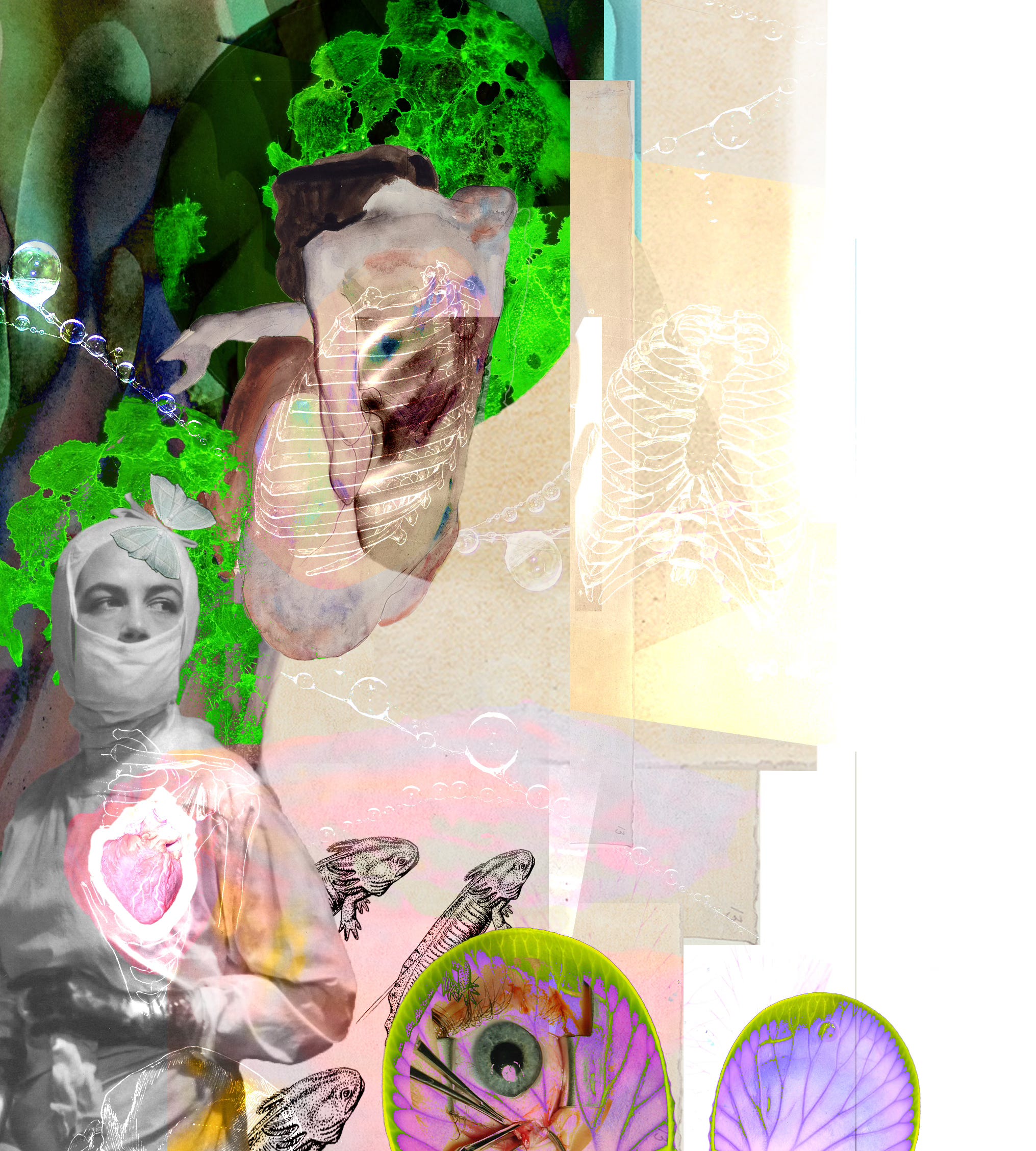
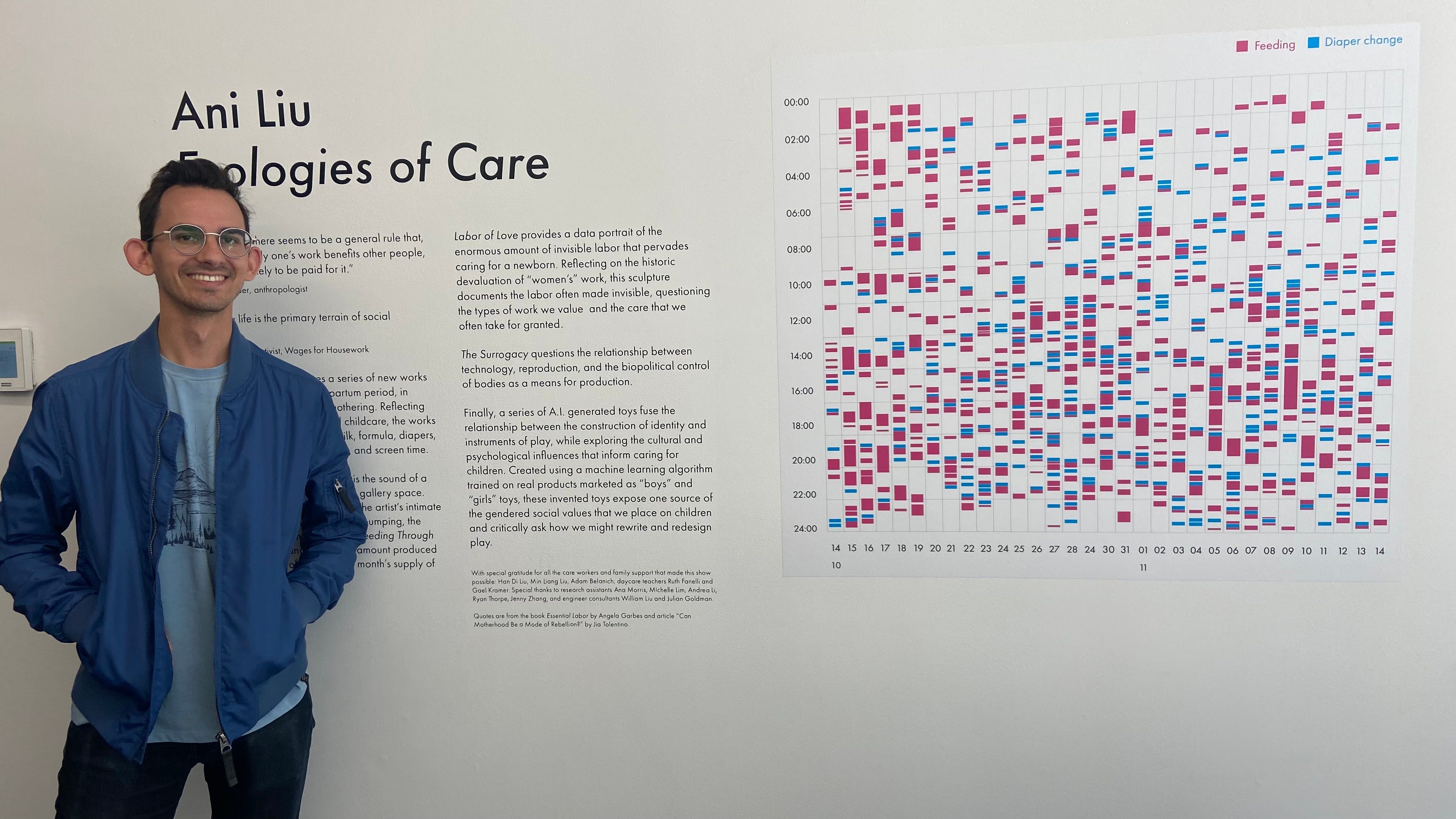
In a small gallery on the Lower East Side, three gallons of artificial breast milk pumped through tubes coiled and scattered across the concrete floor accompanied by a low mechanical whirring. The exhibition, Ecologies of Care from the studio and life of artist Ani Liu, greeted visitors with the trappings of motherhood in action: glass vials filled with breast milk, baby formula, and diaper remnants, meant to illustrate in real time the constant labor required to properly care for a newborn. And in the center of it all—of the imprints of a hardworking new mom and her machines—stood a gently glowing platform on which a curious glass-like object sat.
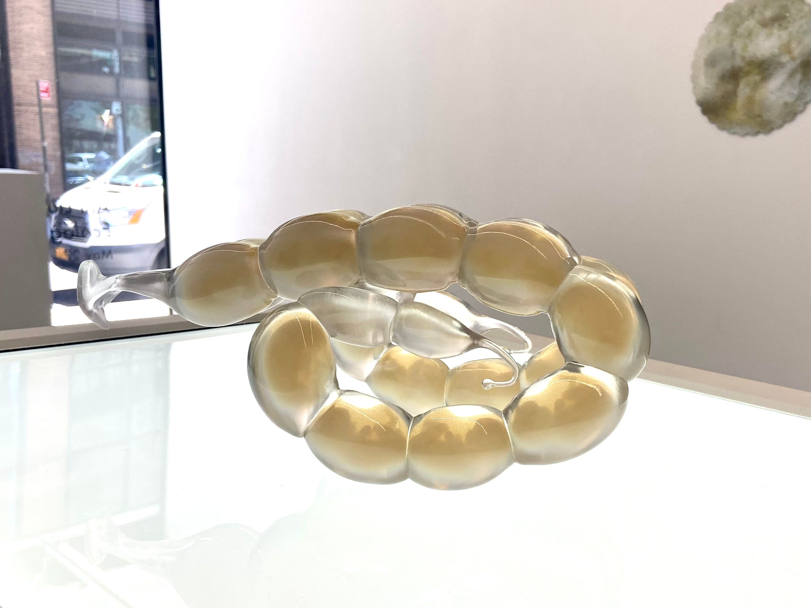
If you’ve ever taken an anatomy class, you may be familiar with what a pig uterus looks like (I will never forget my AP biology teacher’s startled scream as she discovered one floating in a bucket of formaldehyde she forgot she had). The uterus is long and sprawling, and each pig fetus inhabits an individual compartment, stacked along a two-tailed tract. I found myself once again greeted by its undulating form many years after holding one in AP biology. Because on that glowing platform in that small gallery on the Lower East Side sat a precise replica, 3D-printed in a glassy resin, incubating growing pig and human fetuses in chambers side-by-side. That’s right. Pig AND human fetuses. Together.
Believe it or not, Liu based this sci-fi sculpture, The Surrogacy (bodies are not factories), on real-world research. Sometimes science is indeed stranger than fiction, and the field of interspecies pregnancy most certainly toes the line! In a desperate effort to help endangered giant pandas procreate more successfully, scientists in the early 2000’s attempted to impregnate cats with panda embryos. Many of these embryos—which the scientists previously cloned in rabbits—seemed to take, but the project didn’t end up continuing.
While using science to bolster the reproductive rate of an endangered species seems noble, Liu asked another, more extreme question. From pandas to people, what would happen if we tried to do the same thing with human embryos? Unloading the burden of motherhood off women and onto animals?
“In using the pig as a vessel, it felt almost like equating my own body with that of livestock,” Liu said in an interview with Artnet. “I wanted to show that we don’t actually need artificial wombs, we really need better policies.” As the title of Liu’s piece clearly states, Bodies (women’s and pigs included) are not factories, and they (as well as the people they belong to), shouldn’t be reduced to being treated as biological machines. It’s a statement as political as it is scientific.
But what does this have to do with organ transplantation? The Surrogacy offers a critique not only on the social landscape surrounding women’s bodies and our culture of motherhood, but it also raises questions about how we should be using livestock to produce life-sustaining goods.
The pig kidney Richard Slayman received was the product of 69 genetic edits made using CRISPR-Cas9—grown in a living, breathing animal. These edits disrupted (or “knocked down”) three genes involved in the production of antigens that normally activate the human immune system, inserted seven human transgenes into the pig’s embryo to improve integration of the kidney with the patient’s native bloodstream and to reduce inflammation overall, and made 59 additional genetic changes to inactivate retroviruses native to the pig genome. These retroviruses have the potential to infect human cells, potentially creating tumors and immunodeficiencies in the recipient, if active.
Dr. Leonardo V. Reilly, Medical Director for Kidney Transplantation at Massachusetts General, who performed Slayman’s kidney transplant, views these pig kidneys as a way to extend the lifespan of critically-ill patients on the organ transplant list—an option previously off the table for many due to our country’s chronic organ shortage.
Doctor’s from the University of Maryland have similar hopes. In 2022 and 2023, they conducted two heart transplantations using similarly genetically-edited pig organs. The first patient, David Bennett, passed away two months after his procedure in which a latent retrovirus initiated a deleterious inflammatory cascade (alongside a number of other factors) that led to a stiffening of the heart’s muscle tissue and subsequent heart failure.
The second patient, Lawrence Faucette, passed away six weeks after his operation when signs of organ rejection became evident.
However, unlike heart transplant recipients, who unfortunately die if the organ fails, kidney transplant recipients aren’t subject to the same high stakes, as Dr. Reilly succinctly described to Wired:
“If Slayman’s kidney did stop working, he could resume dialysis. The pig heart recipients had no back-up options. Even if pig organs aren’t a long-term alternative, they could provide a bridge to transplant for patients like Layman who would otherwise spend years suffering on dialysis.”
In other words, even if xenotransplants don’t fully succeed at restoring organ function long-term, they may be able to prolong the lives of patients on the organ transplant waiting list until something more permanent becomes available. I can see this as a very plausible success story for genetically-modified xenotransplants. One that balances patient’s needs for timeliness with permanence.
But not everyone sees CRISPRed pig kidneys as the solution to our country’s organ shortage. As reported by The New York Times in late March, Kathy Guillermo, Senior Vice President at People for the Ethical Treatment of Animals, shared her dismay, saying “Researchers need to focus on cleaning up the organ donation system and leave the animals alone.”
And I kind of agree with her—at least with the idea that pig organs may not be the ultimate solution here.
Currently, the United States operates with an opt-in organ donation system, meaning people must definitively express their consent to be organ donors. Switching to an opt-out organ donation system, in which individuals must explicitly express their dissent, is commonly believed to increase rates of organ donation, and parts of the United Kingdom and Canada have switched their systems from opt-in to opt-out with the hopes of accomplishing just that. However, studies show that countries with opt-out programs actually experience a decrease in donations from the living without any significant increase in donations from the deceased.
So if switching to opt-out organ donation in the U.S. isn’t the answer, what is?
I don’t really know. I don’t think anyone really does—hence the pig organs. But one thing definitely unsettles me in all this. The production of these genetically-modified porcine organs at scale would require the mass slaughter of animals for this one specific purpose. It all feels like an unnecessarily convoluted method, one that stretches the burden on our planet—increasing our feedstock requirements, water consumption, land use, and carbon emissions—as well as our morals. But then, I’m not on the organ transplant waitlist.
I’m ultimately deeply conflicted. On one hand, both pigs and cows are already systematically reared to manufacture biomedical products. As Ani Liu reminds us, their bodies are indeed used as factories. Bovine blood is used to produce key cell culture additives, including the much written about fetal bovine serum. The bodies of pigs are harvested for gelatin, heparin, and insulin, among other compounds. Both bovine and porcine heart valves and cartilage tissues are used in valve replacement surgeries. To date, over 1 million people have been treated with the popular Edwards Lifesciences’ SAPIEN transcatheter heart valves, which employ bovine pericardium tissue in their design.
On the other hand, reports from the USDA tell us that 128 million pigs and 28 million cows were slaughtered in 2023 as part of our commercial food system. It seems to me, that the yearly number of animals required by the medical system (hundreds of thousands, perhaps more) pales in comparison to the number demanded by our current food system (hundreds of millions). Surely there is room to be made for these life-saving organs to be produced while minimizing the amount of animals needed in other industries, not only for the well-being of the animals but also for the well-being of our planet, as Jan Dutkiewicz poignantly writes about for The New Republic.
As our field continues to develop, implant, and test genetically-modified organs grown in pigs, like Slayman’s kidney, we’ll have to wrestle with a host of new questions. Is it ethical to farm thousands of pigs for spare parts? Is extending the lives of people, potentially very briefly, worth extinguishing the lives of other living beings? Are there other, more effective ways of increasing the organ donation rate? Are there other tissue-engineered therapies with fewer trade-offs and longer-lasting results? Are there tissue-engineered therapies that won’t require organ recipients to be immune-suppressed? Is this method of manufacturing organs sustainable for both people and planet as the threat of climate change and zoonotic disease grows more extreme?
Both Mr. Slayman, who was released from the hospital two weeks after his kidney transplant, and Mrs. Pisano remain alive and well, their new pig kidneys functioning as they ought.
Until next time,
Matthew
Have news you want to share with the Fleshy Futures community, leave it in the comments below!
Matthew- Thanks for sharing this. I don't know much about tissue engineering, so finding this article is a good find. Hope you're well this week. Cheers, -Thalia